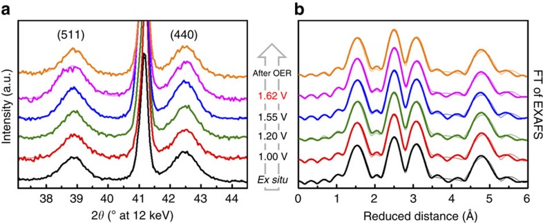
Figure 3. In situ structural characterization of Co3O4 films.
In situ X-ray diffraction patterns (a) and Fourier-transforms (FT) of quasi-in situ EXAFS spectra collected at the Co K-edge (b) of Co3O4 catalyst films. Experimental data and fitted profiles are shown in bold and thin lines, respectively. The diffraction patterns were recorded using grazing-incident excitation at α=0.3° and 12 keV. The electrode potential was increased stepwise from +1.0 to +1.62 V versus reversible hydrogen electrode, the latter representing the catalytically active catalyst state, in 0.1 M KPi at pH 7 (cf. Fig. 2a and Supplementary Fig. 12). The state after OER is a dry state for which the electrode was removed from the electrolyte at +1.0 V rinsed with de-ionized water and dried in N2 flow. The Miller indices of selected Co3O4 reflections are indicated; the diffraction patterns were background corrected for better visualization. Fitting of the diffraction pattern was performed using pseudo-Voigt profiles. Samples for XAS were freeze-quenched under potential control using liquid N2 after 15 min at 1.62 V in 0.1 M KPi. Further details on data analysis are given in the caption of Supplementary Tables 1 and 2. See also Supplementary Figs 15 and 16.

