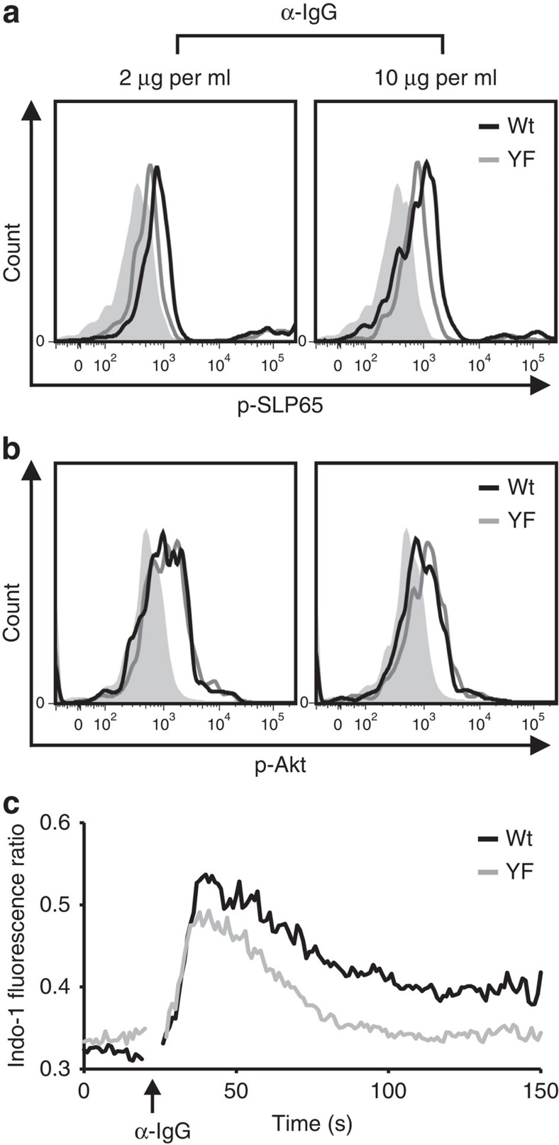
Figure 1. The ITT amplifies BCR-induced Ca2+ signalling.
Splenic B cells from naive wild-type (wt) and ITT-mutant (YF) mice were isolated by magnetic cell sorting and stimulated with either low (2 μg ml−1, left panels) or high (10 μg ml−1, right panels) doses of anti-IgG F(ab')2 fragments. Unstimulated cells served as controls (filled histograms). Subsequently, cells were stained with phospho-specific antibodies to either SLP65 (a) or Akt (b) wt: (black curves), YF: mIgG1-YF (grey curves). (c) Splenic B cells from three age-matched male mice of each genotype that had been immunized with sheep red blood cells for 11 days were pooled, loaded with Indo-1-AM and stained with FITC-labeled monomeric Fab fragments against IgG1. Ca2+ mobilization of mIgG1-positive cells was monitored before and after stimulation with anti-IgG F(ab')2 fragments (20 μg ml−1, indicated by an upward arrow) in the presence of 1 mM extracellular CaCl2. Data are representative of three to four experiments.

