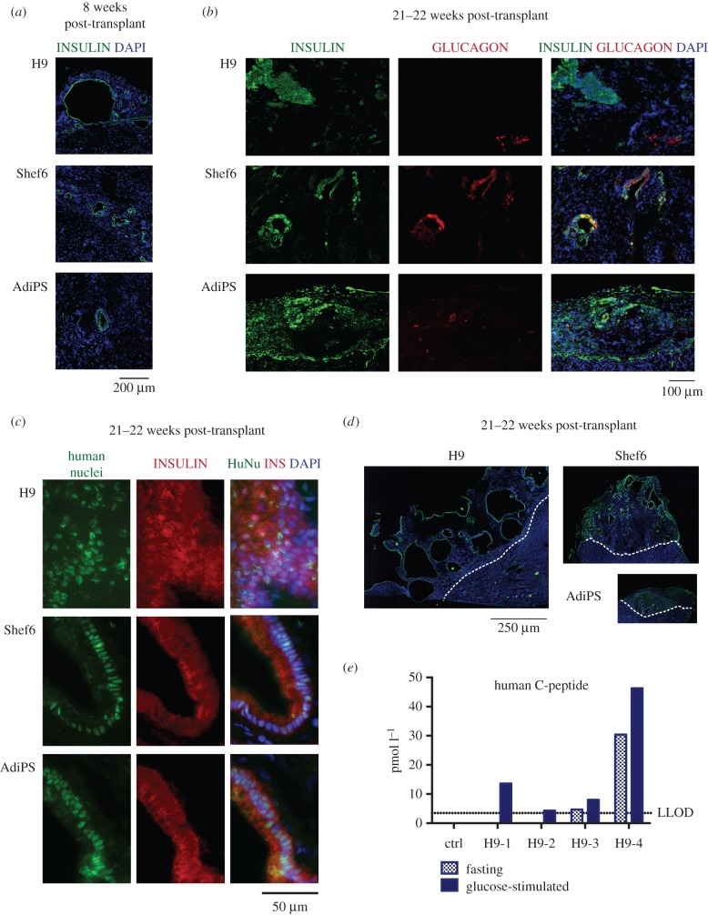
Figure 1.
In vivo maturation of presumptive pancreatic endoderm generated from PSC according to Kroon et al. [3]. (a) Immunostaining for insulin of grafts eight weeks post-transplant derived from differentiated H9, Shef9 and AdiPS. (b) Immunostaining for insulin and glucagon, or (c) human nuclei of grafts 21–22 weeks post-transplant. (d) Immunostaining for insulin of grafts obtained 21–22 weeks after transplanting; representative images showing transversal sections of whole grafts. The images are tiled from multiple fields of view, scale bar for all 250 µm. (e) Glucose-induced C-peptide release in mice engrafted with H9-generated cells. Dotted line indicates lower limit of detection (3.48 pmol l−1), as defined by standards measurements.

