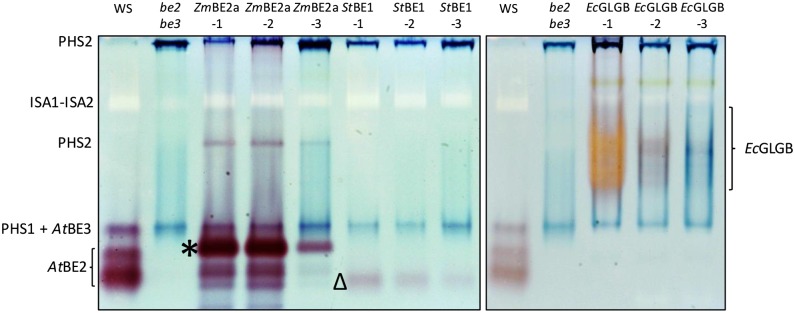
Figure 3.
Native PAGE (zymograms) of be2be3 mutants expressing different BE types. Soluble proteins were extracted from the indicated lines and separated by native PAGE in gels containing 0.02% (w/v) glycogen. BE activity was detected by incubating the gels in a medium containing phosphorylase-a and its substrate, Glc-1-P. The identifications of the endogenous BE2 and BE3 activities are based on comparable analysis of the wild type, the be2 and be3 single mutants, and the be2be3 double mutant (see supplemental data in Pfister et al., 2014). Dark bands represent the endogenous cytosolic (PHS2) and plastidial (PHS1) α-glucan phosphorylases. In these gels, BE3 comigrates with PHS1. A pale band corresponds to the heteromultimeric ISA1-ISA2 DBE. Unique activities attributable to ZmBE2a (*), StBE1 (Δ), and EcGLGB are indicated.

