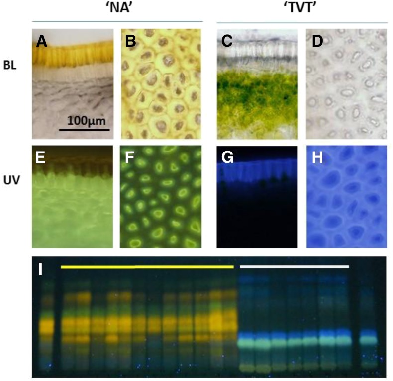
Figure 1.
DPBA staining of muskmelon fruit rinds. Microscopic view of fruit rinds of the parental lines NA (A, B, E, and F) and TVT (C, D, G, and H). A, C, E, and G are fruit rind cross sections, and B, D, F, and H are upper views of fruit rind. BL, Bright visible light. I, TLC of methanol extract stained with DPBA of NA (left lane) and TVT (right lane) marked by yellow and white lines are the 12 F3 yellow families and 7 F3 white families, respectively.

