Figure 6.
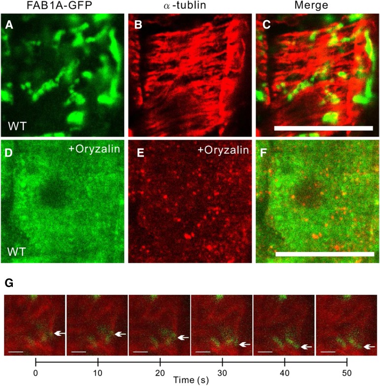
The structure of FAB1-positive endosomes is impaired by oryzalin treatment. Immunofluorescence detection of FAB1A-GFP and α-tubulin in the wild type (WT; A–C) and wild type with oryzalin treatment (D–F). G, Time lapse series of mRFP-β-tubilin (TUB6)-labeled microtubules (MTs; red) and FAB1A-GFP-positive endosomes (green). Arrows depict clustered endosomes along with cortical microtubules. (See also Supplemental Movie S1.) Green and red signals represent FAB1A-GFP and β-tubilin, respectively.

