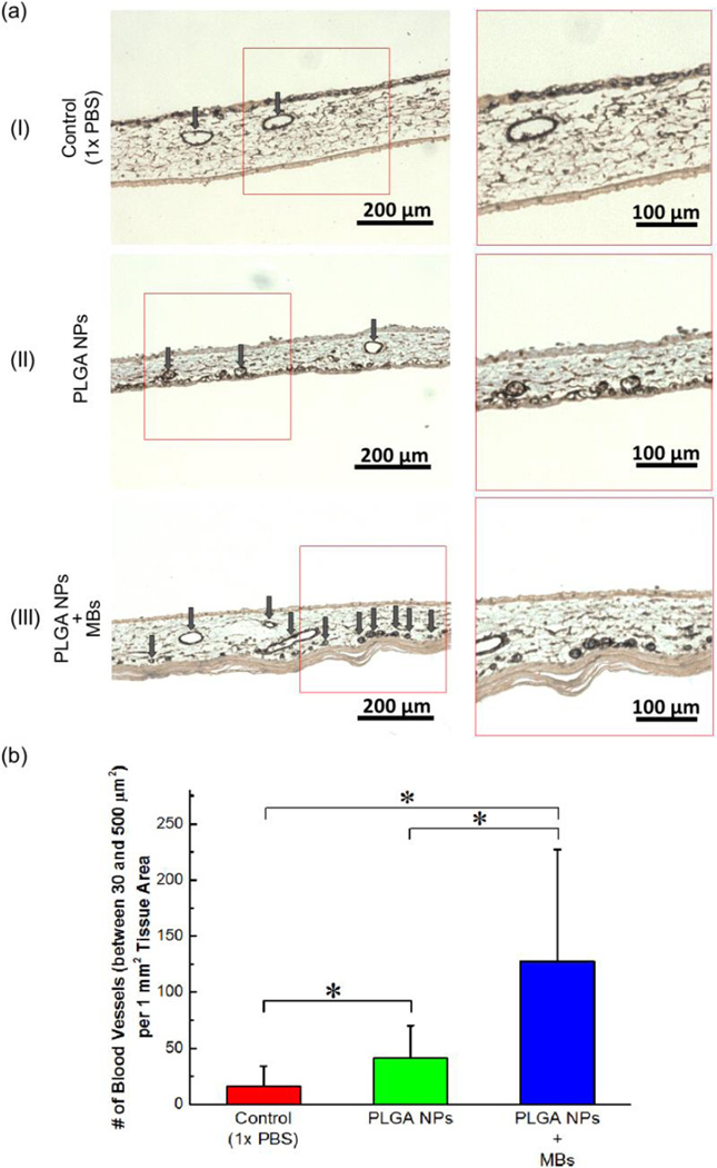
Figure 4.
In vivo evaluation of vascularization stimulated by Ang1-encapsulated PLGA nanoparticles (NPs). (a) Histological images of the cross session of CAMs stained for α-SMA. (a-I) CAM treated with PBS, (a-II) CAM treated with Ang1-encapsulated PLGA NPs, and (a-III) CAM treated with Ang1-encapsulated core-shell particles in which PLGA NPs were immobilized on the microbubbles (MBs). The images on the right column represent magnified views of the boxed region in the images on the left column. Arrows indicate mature blood vessels positively stained for α-SMA, marked by brown color. (b) Quantified number of mature blood vessels with cross-sectional area between 30 and 500 µm2 per tissue area of 1 mm2. The values and error bars represent average values and standard deviation of 9 samples per condition. * represents the statistical significance of the values between conditions (* p < 0.05).

