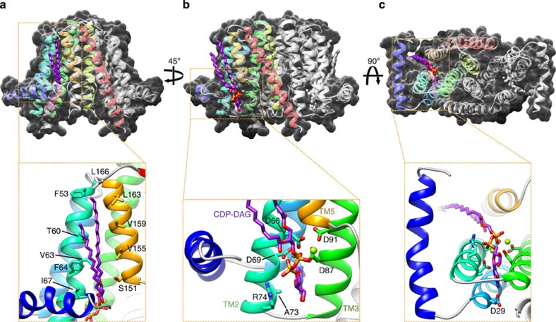
Figure 3. CDP-DAG binding to RsPIPS-FL.
The structure of RsPIPS-FL, depicted in ribbon representation and coloured as per Fig. 1, is shown in three different views (a–c) superimposed on a transparent ‘Stromboli black' spacefill model (upper panels), with matching magnified insets of boxed regions below (lower panels). CDP-DAG is depicted in purple and Mg2+ ions in light green. Side chains that contact CDP-DAG or Mg2+ are shown in stick representation.

