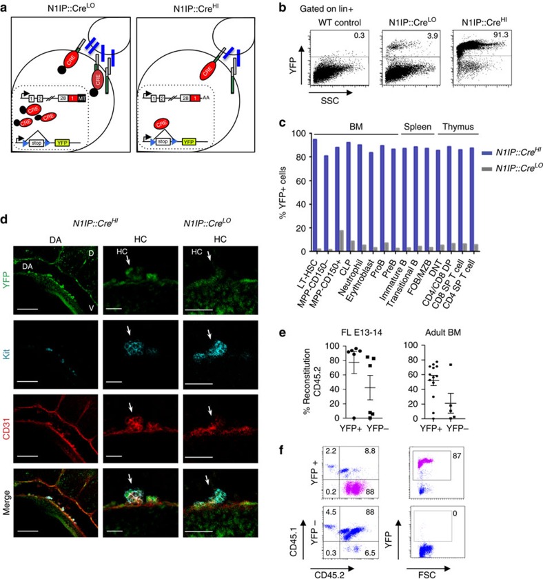
Figure 1. Haematopoietic and arterial specification requires different levels of Notch1 activity.
(a) Schematic representation of Notch activation history mouse reporters by replacing the intracellular domain of mouse Notch1 with low sensitivity (N1IP::CreLO) and high sensitivity (N1IP::CreHI) Cre-recombinase. Reporter activation of N1IP::CreLO requires a high threshold of Notch activity, while N1IP::CreHI is induced in response to low or high Notch activity. (b) Flow cytometry analysis of peripheral blood of adult mice. Cells were stained with Lineage (lin) markers (CD3, B220, Gr1, Mac1 and Ter119) gated on lin+ cells. Numbers indicate the percentage of YPF+ cells. (c) Graph represents the percentage of YFP+ cells within haematopoietic cell types in the bone marrow (BM), spleen and thymus of N1IP::CreLO (grey bars) and N1IP::CreHI (blue bars) as detected using flow cytometry. (d) Representative confocal images of three-dimensional whole-mount immunostaining in N1IP::CreHI and N1IP::CreLO embryos (E10.5) detecting YFP (green), c-Kit (cyan) and CD31 (red). General view of the dorsal aorta (left panel) and details of haematopoietic cluster (right panels). White arrows indicate cluster structures. D, dorsal; DA, dorsal aorta, HC, haematopoietic cluster; V, ventral. Scale bars, 100 μm for DA, 25 μm for HC in N1IP::CreHI and 50 μm in N1IP::CreLow. See also Supplementary Fig. 1. (e,f) Graphs show the percentage of reconstituted cells in animals transplanted with YFP+ and YFP− fractions of E13-14 fetal liver and BM at 4-month post-transplantation (e). Representative dot plots from analysis (f). Donor CD45.2 N1IP::CreHI cell fractions together with 500,000 supporting CD45.1 spleen cells were transplanted into CD45.1/CD45.2 chimeras.

