
Figure 6.
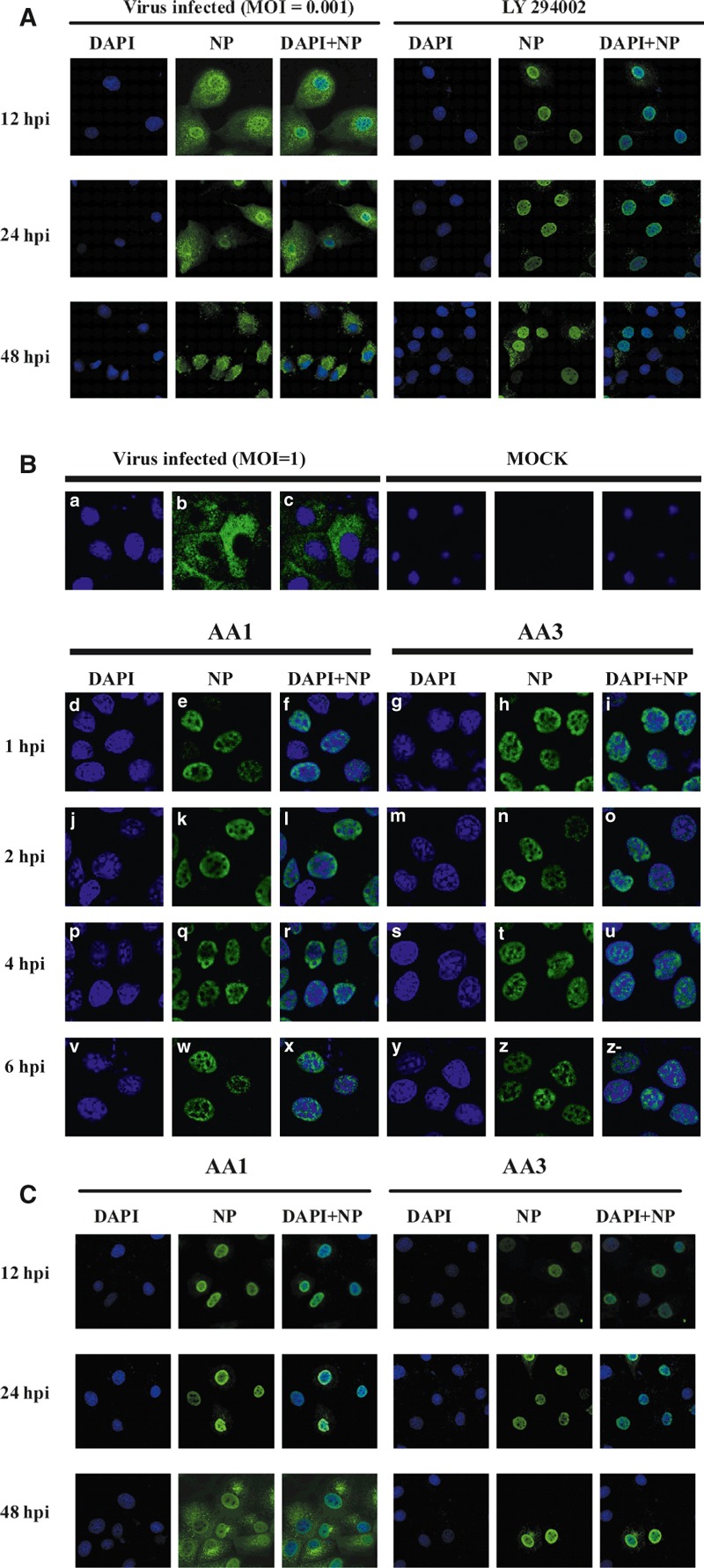
178 H5N1 HPAIV (MOI = 1) and MDCK cells were either untreated or were treated with AA1 or AA3 as follows: (−) AA, normal infection with no AA treatment as control (Figure 6A,B); The location of NP was tested in single replication (8 h p i), cells treated with AA1 (4·67 μm), AA3 (4·90 μm) after 1 h of virus adsorption at 4°C. Infected cells were then incubated in medium with AA1 (d, e, f, j, k, l, p, q, r, v, w, x) or AA3 (g, h, I, m, n, o, s, t, u, y, z, z‐) at different time intervals (1, 2, 4 and 6 h p.i.) until 8 h p.i., and the intracellular amount and localization of viral RNPs (green, e, h, k, n, q, t, w, z), as well as the nuclei (blue, d, g, j, m, p, s, v, y), DAPI + NP (f, I, l, o, r, u, x, z‐) were detected by immunofluorescence. The location of NP protein at muti‐round replication (24 and 48 h p i) was tested in Figure 6C.