
Fig 3. DNA binding domain of bacteriophage repressors.
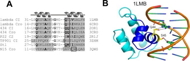
(A) Sequence alignment of the HTH motifs of bacteriophage repressors and Stl. The number before each segment is the amino acid sequence number of the first residue in the sequence. Helices are with gray background, similar residues are in bold, box highlights residues interacting with DNA nucleobases. PDB ID of the proteins is indicated on the right side of the sequences. (B) Experimentally determined structure of the DNA-bound CI bacteriophage repressor DNA cartoons orange, protein cartoon: dark blue for HTHs, otherwise cyan. DNA bases and DNA interacting amino acid residues are stick representation with atomic coloring (protein carbon yellow, DNA carbon green, oxygen red, nitrogen blue, phosphorus orange.) The PDB ID of the structure is indicated. Stereo representation of all experimentally determined protein-DNA complex structures represented in the sequence alignment in this figure are available in S3–S9 Figs.