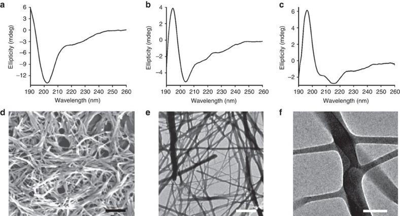
Figure 2. Structural analysis and self-assembly of Aib containing peptides.
CD spectra of the designed peptides: (a) SHR-LL, (b) SHR-FL and (c) SHR-FF. (d) SEM micrograph of the peptides SHR-FF. Scale bar, 2 μm. (e) TEM micrograph of the peptides SHR-FF. Scale bar, 1 μm. (f) High resolution TEM images showing the presence of branching along the length of the fibres. Scale bar, 100 nm.

