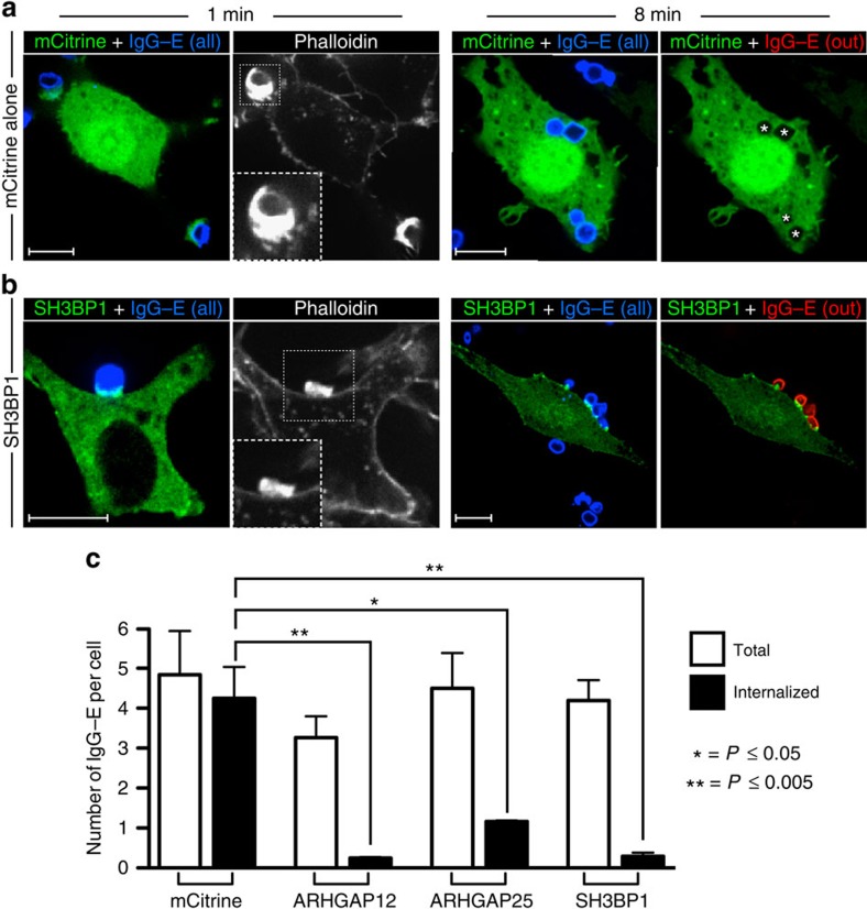
Figure 4. Functional validation of candidate RhoGAPs.
(a,b) Constructs encoding mCitrine alone (a) or SH3BP1 (b) were electroporated into RAW 264.7 macrophages to yield high levels of expression. To determine whether these RhoGAPs could indeed inactivate GTPases that are instrumental for phagocytosis, electroporated cells were challenged with IgG-opsonized erythrocytes and allowed to interact with the targets for 1 min (left panels) or 8 min (right panels) before fixation. All phagocytic targets (shown in blue) were stained with a Cy5-conjugated anti-IgG secondary antibody before addition to phagocytes. To determine the number of IgG-coated erythrocytes that were not internalized, fixed (non-permeabilized) cells were stained with Cy3-conjugated anti-IgG (shown in red). Cells were then washed, permeabilized and stained for F-actin with phalloidin. Insets show magnified views of nascent phagocytic cups (boxed region) at the 1 min time point. Phagosomes that had already been formed before addition of the Cy3-conjugated secondary antibody are indicated with a star. Scale bar, 10 μm. Micrographs are representative of three independent experiments. (c) Quantification of the total number of IgG-coated erythrocytes that were engaged (white bars) or internalized (black bars) per phagocyte overexpressing mCitrine alone or mCitrine-tagged ARHGAP12, ARHGAP25 or SH3BP1. Values in (c) represent the means of three independent replicates±s.e.m. *P≤0.05 and **P≤0.005 using Student's two-tailed unpaired t-tests. At least 20 cells were assessed per replicate for each of the RhoGAPs examined.

