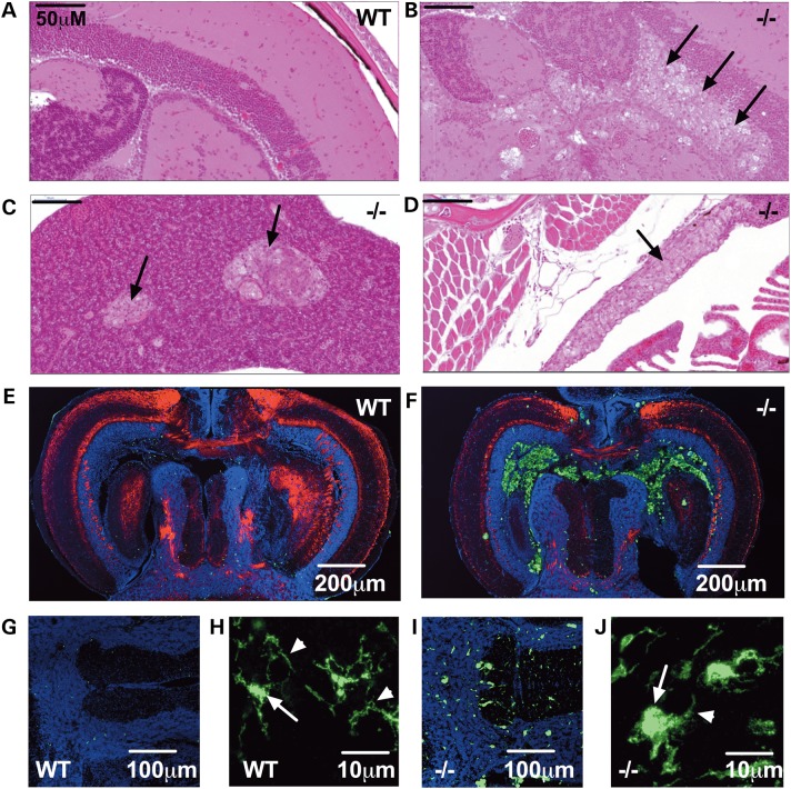
Figure 5.
gba1 deficiency leads to Gaucher cell invasion and increased abundance of activated microglia in gba1−/− brain. H&E staining of 12 wpf sections demonstrated Gaucher cell (black arrows) organ invasion of the tectal ventricle within the brain of gba1−/− (B), compared with wild-type (WT) brains (A). Gaucher cell organ invasion was also present in the visceral organs of gba1−/− such as the liver (C) and thymus (D). Fluorescent images show confocal micrographs of gba1−/− and wild-type siblings control brains labeled by indirect immunofluorescence for 4.C4 (macrophages and microglia; green), DAPI (nuclei; blue) and P0 (myelin; red). Low-power images through the tectal ventricle showed accumulation of 4.C4 immunoreactive macrophages in the ventricle and periventricular region of gba1−/− (F) but not wild-type brain (E). Microglia within the brain parenchyma were identified by their immunoreactivity to 4.C4 and typical morphology (G and I). Compared with wild-type brain (G), microglial were more numerous and brightly labeled in gba1−/− brain (I). In addition, compared with the typical quiescent morphology of microglia seen in wild-type brain (H), marked rounding of the cell body and retraction of processes was apparent in gba1−/− brain (J). The white arrows point at a normal microglial cell body in a wild-type control brain (H) and a rounded microglial cell body in a gba1−/− brain (J); the white arrow heads point at a normal, extended microglial process in a wild-type control brain (H) and at a retracted microglial process in a gba1−/− brain (J). These morphological changes are typical of microglial activation.

