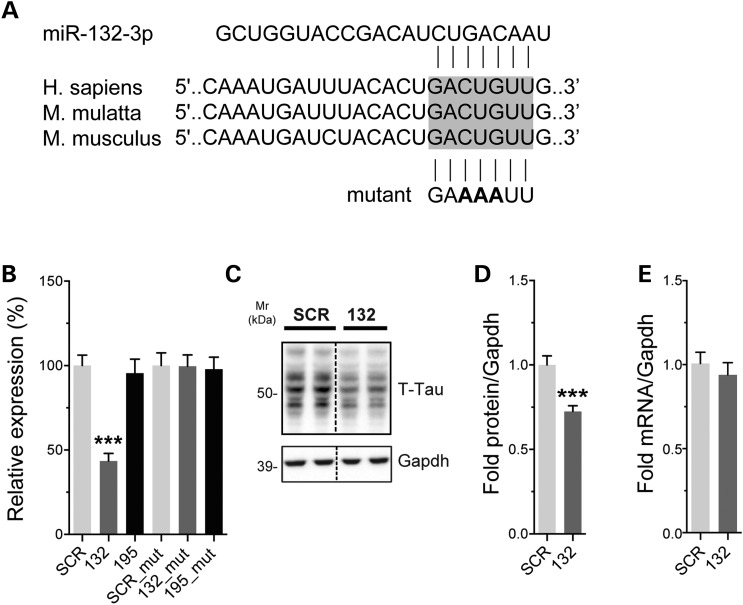
Figure 2.
Tau is a direct target of miR-132. (A) Sequence of the tau 3′ UTR showing the predicted miR-132/212-binding site. This region is highly conserved (shown here is human, monkey and mouse sequences). The miR-132 site mutation is shown in bold. (B) Left panel: Neuro2a cells were transfected with 50 nm final concentration of miR-132 or miR-195 mimics. Twenty-four hours post-transfection, luciferase signal was measured. Signals were normalized to Renilla luminescence for transfection efficiency, and graph represents the relative luciferase signals compared with a scrambled mimic control (SCR). Right panel: luciferase assays were performed using a mutant 3′ UTR construct for tau. Here, cells were treated with and 10 nm of miR-132 or miR-195 mimics. The tau mutation completely blocked the effects of miR-132. (C, D) Representative western blot of naive Neuro2a cells treated with 50 nm final concentration of miR-132 or SCR control. Cells were lysed 24 h post-transfection. Gapdh was used as normalization control, and quantifications are shown (n = 3 in triplicate). (E) Tau mRNA quantification after miR-132 overexpression (50 nm) in Neuro2a cells compared with SCR control. Gapdh served as normalization control (n = 2 in triplicate). Statistical significance was assessed by one-way ANOVA with Bonferroni multiple comparison test, where ***P < 0.001 and by Student's unpaired t-test, where ***P < 0.001.

