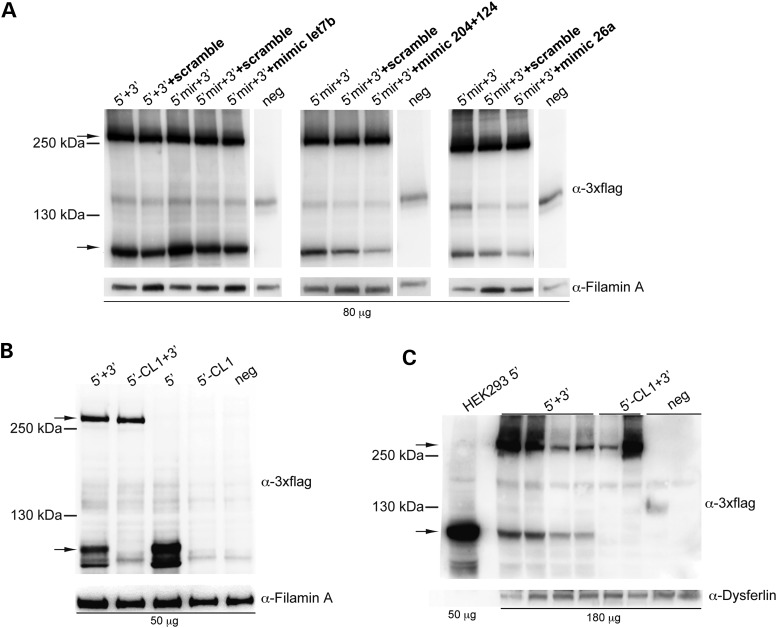
Figure 4.
Inclusion of the CL1 degradation signal but not of miR target sites in the 5′-half vectors results in significant reduction of truncated proteins. (A) Representative Western blot analysis of HEK293 cells infected with dual AAV2/2 hybrid vectors encoding for ABCA4, containing miR target sites for either miR-let7b (4×, left panel), miR-204 + 124 (3×, central panel) or miR-26a (4×, right panel). 5′ + 3′: cells co-infected with 5′-half vectors without miR target sites and 3′-half vectors; 5′mir + 3′: cells co-infected with 5′-half vectors containing miR target sites and 3′-half vectors; neg: control cells infected with the 3′-half vectors; +scramble: cells infected in the presence of scramble miR mimics; +mimic let7b: cells infected in the presence of miR-let7b mimics; +mimic 204+124: cells infected in the presence of miR-204 and -124 mimics; +mimic 26a: cells infected in the presence of miR-26a mimics. (B and C) Representative Western blot analysis of either HEK293 cells infected with dual AAV2/2 hybrid vectors (B) or pig eyes (c; RPE+retina) 1-month post-injection of dual AAV2/8 hybrid vectors encoding for ABCA4 and containing or not the CL1 degradation signal. 5′ + 3′: cells co-infected or eyes co-injected with 5′-half vectors without CL1 and 3′-half vectors; 5′-CL1 + 3′: cells co-infected or eyes co-injected with 5′-half vectors containing CL1 and 3′-half vectors; 5′: cells infected with 5′-half vectors without CL1; 5′-CL1: cells infected with 5′-half vectors containing CL1; neg: control cells infected or control eyes injected with either the 3′-half vectors or EGFP-expressing vectors, as negative controls. (A–C) The upper arrows indicate full-length ABCA4 proteins, the lower arrows indicate truncated proteins; the molecular weight ladder is depicted on the left. The micrograms of proteins loaded are depicted below the image. α-3×flag: Western blot with anti-3×flag antibodies; α-Filamin A: Western blot with anti-Filamin A antibodies, used as loading control; α-Dysferlin: Western blot with anti-Dysferlin antibodies, used as loading control. (A and B) The Western blot images are representative of n = 3 independent experiments. (C) The Western blot image is representative of n = 5 eyes injected with 5′ + 3′ vectors, n = 2 eyes injected with 5′-CL1 + 3′ vectors and n = 5 of eyes injected with either the 3′-half vectors or EGFP-expressing vectors as negative controls.

