Fig. 2.

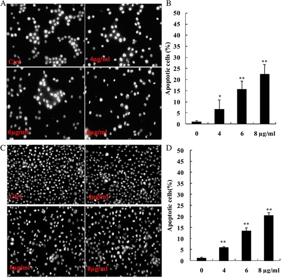
DAPI stained apoptotic cells in morusin treated MCF-7 and MDA-MB-231 breast cancer cells. a DAPI stained apoptotic cells in morusin treated MCF-7 cells. b The histogram shows that there was significant increase of AO/EB stained apoptotic cells in morusin treated MCF-7 cells. c DAPI stained apoptotic cells in morusin treated MDA-MB-231 cells. d The histogram shows that there was significant increase of DAPI stained apoptotic cells in morusin treated MDA-MB-231 cells. *P < 0.05, **P < 0.01. Three independent experiments were performed
