Fig. 4.

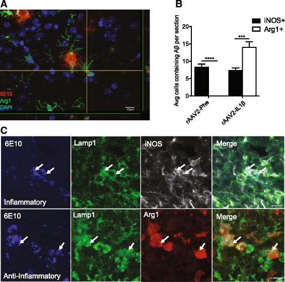
Aβ is taken up by Arg1+ microglia. a Representative confocal images of Arg1+ cell (green) containing Aβ (red) after rAAV2-IL1β injection. DAPI (blue) is used as to counter stain cell nuclei. Scale bars represent 10 μm. b Quantification of iNOS+ or Arg1+ microglia in the hippocampus that contain Aβ after 4 weeks rAAV2-Phe or rAAV2-IL1β injection. Data displayed as mean ± SEM, n = 5 animals, ***p < 0.0001, ****p < 0.00001. c Representative images of Arg1+ or iNOS+ microglia containing 6E10 inside the lysosome (Lamp1). Scale bars represent 10 μm
