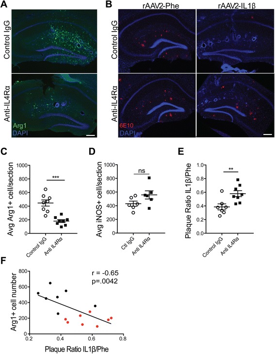
Fig. 7.

IL-4 signal blockade inhibits Arg1+ microglia induction and partially impairs Aβ plaque reduction. Representative images depict Arg1+ microglia (a, green) and 6E10 (b, red) 28 days post-injection and cannula implantation. Scale bar represents 100 μm. Quantification of Arg1+ cell number (c), iNOS+ cell number (d), and 6E10 Aβ plaque area (e), in the hippocampus. Data was analyzed with Student’s t test. n = 7–8 animals; mean ± SEM shown; **p < 0.005, ***p < 0.0001. f Correlation was determined by plotting the Arg1 cell count by plaque ratio for each animal. The plaque ratio was generated by dividing the amount of plaque area in the inflamed hemisphere by the amount of plaque area in the control hemisphere. This was performed on three hippocampal sections per mouse then averaged together. The higher the number, the more plaque is present in the inflamed hippocampus. Each dot represents one animal. Black dots denote the control IgG while red dots denote mice that received anti-IL-4Rα. r Pearson’s coefficient
