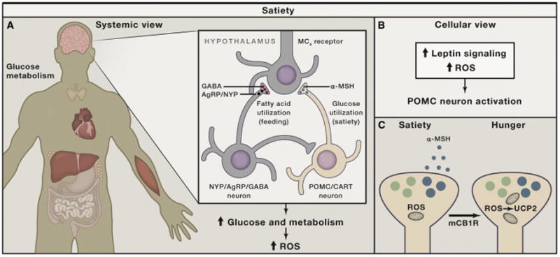
Figure 3. Schematic Illustration of Hypothalamic Control of Positive Energy Metabolism with Elevated ROS.

(A) Satiety (feeling full) is promoted by hypothalamic neurons (blue) that produce pro-opiomelanocortin (POMC)-derived peptides, such as α-MSH, which, in turn, act on melanocortin-4-receptor-containing neurons. When these neurons are active, systemic metabolism is shifting toward glucose utilization, with enhanced mtROS production contributing to increased cellular ROS in various tissues.
(B) The activation of POMC neurons after a meal is accomplished by ROS, in part driven by increased mtROS production, and is supported by intracellular leptin (Jak/Stat) and insulin (PI-3K/PTEN) signaling, involving altered K-ATP channel activity.
(C) Recent studies indicate that, under unique circumstances (e.g., activation of cannabinoid receptors), POMC neurons, while still driven by ROS, will become promoters of hunger rather than satiety because they shift to releasing appetite-stimulating opiates (β-endorphin) though UCP2-dependent mitochondrial adaptations.
