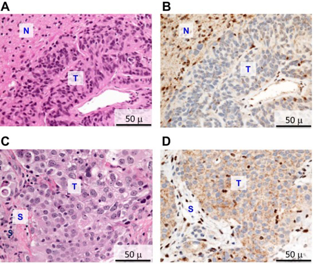
Figure 3.

H&E and immunohistochemical staining of meningioma and mesothelioma tissues from patient III-10. A) H&E staining of meningioma. B) BAP1 immunohistochemistry of meningioma, showing absence of nuclear BAP1 staining in tumor (T) and both cytoplasmic and strong nuclear staining in adjacent normal brain parenchyma (N). C) H&E staining of peritoneal mesothelioma. D) BAP1 immunohistochemistry of same mesothelioma, showing absence of nuclear BAP1 staining in tumor (T) and both cytoplasmic and strong nuclear staining in normal stroma (S).
