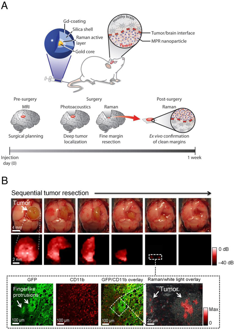
Figure 3.
Multimodal SERS nanoparticles for pre- and intraoperative imaging of malignant brain tumors. (A) Triple-modality nanoparticle imaging concept. Top: the nanoparticle is detectable by SERS, photoacoustic and MR imaging. Nanoparticles are injected intravenously and home to the brain tumor but not to healthy brain tissue. Bottom: because of the stable, long-term internalization of the nanoparticles within tumor tissue, pre-operative MRI for staging and intraoperative imaging with SERS and photoacoustic imaging can be performed with a single injection. (B) SERS-guided brain tumor resection in living mice. Top: intraoperative photographs show the sequential resection steps, and SERS imaging shows the corresponding residual tumor tissue at each resection step. Of note, after gross total resection, there is persistent SERS signal in the normal-appearing resection bed, suggesting the presence of residual cancer (white dashed square). Bottom: subsequent histological analysis of the tissue containing these SERS-positive foci demonstrates residual cancer tissue invading into the surrounding normal brain. Adapted, with permission, from reference (7).

