
Figure 1.
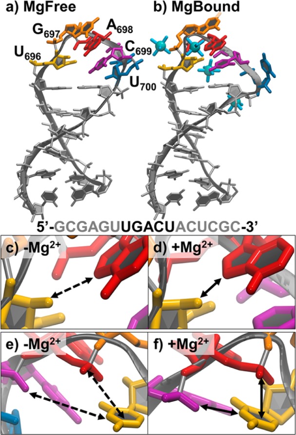
NMR structures and U-turn characteristics of SLV. (a) The MgFree most representative structure determined by NMR (1TBK). Bases of the five-membered U-turn are labeled and shown in color. (b) The MgBound most representative structure determined by NMR (1YN2). Bases of the five-membered U-turn are shown in the same color as part a, and hexahydrated Mn2+ are shown as blue spheres. The primary sequence is shown with the bases of the U-turn in black. Close ups of the characteristic U-turn hydrogen bond between the 2′ OH of U696 (yellow) and the N7 of A698 (red) (c) in the absence of Mg2+ and (d) in the presence of Mg2+. Close ups of the characteristic U-turn hydrogen bond between the 3′ phosphate of A698 (shown in purple here as the 5′ phosphate of C699) and the N3 of U696, and stacking of 5′phosphate of A698 with the U696 base in the (e) absence and (f) presence of Mg2+. Solid lines indicate a favorable interaction (stacking or hydrogen bonding) is present and dashed lines indicate absence of interaction.