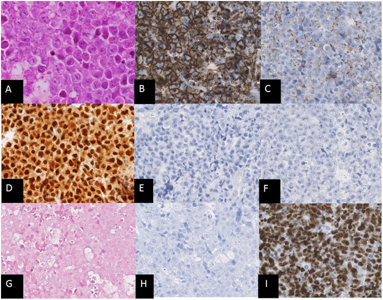
Fig. 2.

a Hematoxylin and eosin staining showing large lymphoid cells with centroblastic morphology (X400). Cells of the oral tumor (X200) were immunohistologically positive for b CD138 and c CD38, d MUM-1, and were negative for e CD20, f CD79a, g EBV-encoded RNA in situ hybridization (EBER-ISH) and h HHV8. i The Ki-67 proliferation index was almost 100 %
