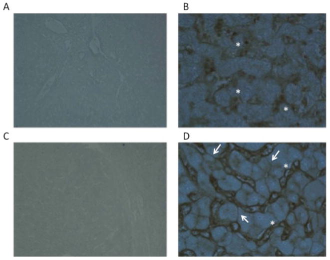
Figure 2.
Lectin staining of HCC or adjacent normal tissue with a recombinant Aleuria aurantia lectin (AAL) that has greater affinity for core fucosylated glycan. Panels A (4X) and B (20x) are from tissue adjacent to the HCC. Areas of staining indicated with the asterisks are the liver sinusoids, which stain with the core fucose binding lectin. Panels C (4X) and D (20x) are from the HCC tissue. In addition to the liver sinusoids, which stain with the core fucose binding lectin as in panels A and B, defined staining of hepatocytes, as indicted by the arrows, can also be seen.

