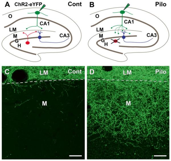
Fig. 12.2.
Axonal reorganization of remaining somatostatin (SOM) neurons in pilocarpine (Pilo)-treated mice at 2 months after status epilepticus, illustrated schematically in (a, b) and in confocal images in (c, d). (a) This schematic illustrates the normal circuitry of SOM neurons in the hilus (red) and s. oriens (green) and the labeling protocol. In a control SOM-Cre mouse, selective labeling of SOM neurons in s. oriens (O) of CA1, by Cre-dependent AAV transfection of ChR2-eYFP, leads to labeling of their axon terminals that are confined to s. lacunosum-moleculare (LM). SOM neurons (red) in the hilus (H) innervate the outer molecular layer (M) of the dentate gyrus where they form synapses with dentate granule cells (G, blue). These hilar SOM neurons are not labeled by the injection in s. oriens. (b) In pilocarpine-treated mice, similar labeling of SOM neurons in s. oriens leads to axonal labeling not only in s. lacunosum-moleculare of CA1 but also in the molecular layer of the dentate gyrus, a region that was previously innervated by vulnerable SOM neurons (red) in the hilus. (c) In a control SOM-Cre mouse, eYFP-labeled axons are concentrated in s. lacunosum-moleculare (LM), and only a limited number of labeled fibers cross the hippocampal fissure (dashed line) to enter the molecular layer (M) of the dentate gyrus. (d) In a similarly transfected pilocarpine-treated mouse, numerous labeled fibers cross the hippocampal fissure and form an extensive plexus in the outer two-thirds of the dentate molecular layer, where they innervate dentate granule cells. Scale bars, 20 μm (Adapted from data in Peng et al. [39])

