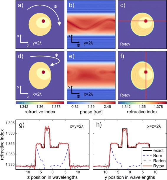
Fig. 2.

Refractive index reconstruction from 3D FDTD simulations. a, d Cross-sectional slices of the 3D cell phantom through the nucleolus located at x=y=z=2 λ from the center of the volume. The color scale is identical to that used in Fig. 1. b, e Sinogram slices matching the cross-sectional position of (a) and (d) for rotational position of ϕ ranging from 0 to 360 degrees; computed with FDTD simulations. c, f Cross-sectional slices of the reconstruction with the Rytov approximation at the same coordinates as in (a) and (d). g, h Line plots through the reconstructed cell phantom with the Born, Radon, and Rytov approximations. The positions of the line plots are shown in (c) and (f). A total of 200 projections were used for the reconstruction
