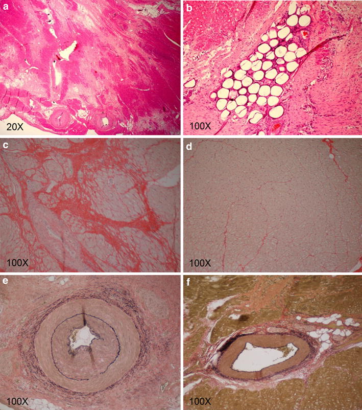
Figure 5.

Histopathology of infarcted versus normal myocardium. Hematoxylin and eosin staining of representative infarcted areas demonstrated a heterogenous area of dense, fibrous, connective tissue intermixed with islets of intact cardiomyocytes (a). Microsphere silhouettes were visualized occluding small arteries and arterioles shown here as stain filling defects (b). Further evaluation with Picro Sirius stain revealed a large amount of disorganized collagen deposition and perifiber fibrosis in the infarcted region denoted by the deeply red staining sections (c), when compared to normal myocardium (d). Evaluation of VVG stained slides demonstrated hyperplasia of the arterial walls, particularly in the media and adventitial layers (e) when compared to normal arteries (f).
