Figure 4.
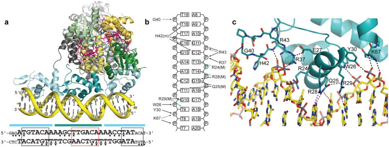
CarH DNA binding. (a) CarH tetramer bound to a 26-bp DNA segment (yellow). CarH is shown in ribbons with one head-to-tail dimer in green (Cbl-binding domains) and yellow (helix bundles) and the other dimer in gray. AdoCbl shown as in Fig. 2a. DNA-binding domains are shown in cyan. Sequence of DNA segment used for crystallization (larger font) as well as flanking sequences in the operator (smaller font) are shown below. Cyan bars indicate base pairs covered by each DNA-binding domain. Base pairs covered by the recognition helix are boxed; red box highlights the promoter −35 element. Nucleotides protected from hydroxyl radical cleavage are indicated by bullets. The orientation of the DNA in the structure was confirmed by heavy atom labeling (Extended Data Figure 6b-d). (b) Schematic of CarH:DNA interactions for the central DNA-binding domain, denoted as follows: black arrows: hydrogen bonds/electrostatic interactions; green arrows: van der Waals interactions; solid lines: contacts from protein side chains; dashed lines: contacts from main chain; (M): contacts in the DNA major groove; (m): contacts in the DNA minor groove. (c) Close-up of interactions between CarH (cyan) and DNA (yellow). Hydrogen bonds and ionic interactions to the phosphate backbone are shown as black dashed lines. Contacts to DNA bases are shown in purple. Side chain orientation is not unambiguous due to the modest resolution of this structure, but many of the contacts shown are supported by mutagenesis (Extended Data Figure 7).

