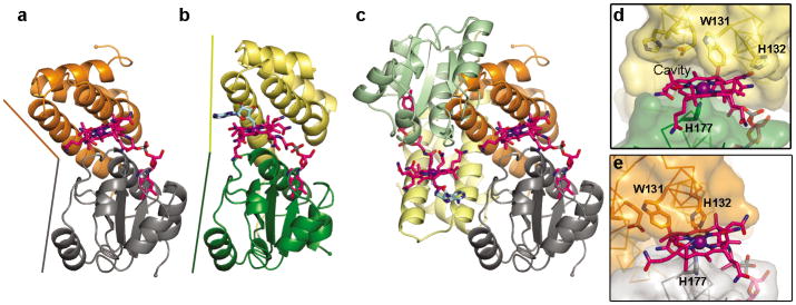
Figure 5.
Light-induced conformational changes in CarH. (a) Structure of light-exposed CarH, with the helix bundle in orange and Cbl-binding domain in gray. DNA-binding domain is hidden for clarity (see Extended Data Figure 8a). Cbl shown with carbons in pink and cobalt in purple. (b) Structure of a CarH protomer in the dark state, shown with helix bundle in yellow, Cbl-binding domain in green, and 5′-dAdo group in cyan. Colored lines highlight domain orientations. (c) Light-induced helix bundle movement causes tetramer disassembly. Shown is a head-to-tail dimer of CarH in the dark state, but the right protomer is replaced by the structure of light-exposed CarH to show the steric clash. (d) Departure of the 5′-dAdo group after light exposure leaves a large cavity on the Cbl upper face. The helix bundle (yellow) and the Cbl-binding domain (green) are shown in surface representation with selected residues shown as sticks. (e) Helix bundle movement fills the cavity at the Cbl upper face and brings His132 to the cobalt, where it occupies the open coordination site. Coloring as in (a).

