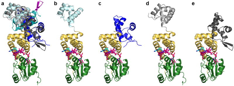
Extended Data Figure 2.
The CarH DNA-binding domain is flexible in the absence of DNA. (a) Overlay of five CarH protomers, including the protomer shown in Figure 2a, highlighting flexibility of DNA-binding domains. Structures are aligned by the Cbl-binding domains (green) and helix bundles (yellow) and shown in the same orientation as Figure 2a. DNA-binding domains are colored in dark cyan, light cyan, dark blue, black, and gray. AdoCbl is shown with Cbl carbons in pink, 5′-dAdo group carbons in cyan, and cobalt in purple. (b-e) Individual CarH protomers shown side by side. Orientation and coloring as in (a).

