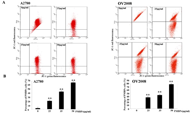
Fig 3. PMBPs induce loss of mitochondrial membrane potential.
(A) The A2780 and OV2008 cells were treated with PMBPs (0, 25, 35, 50 μg/ml) for 24 h and then harvested, stained with JC-1 dye, and finally analyzed by flow cytometry. (B) PMBPs treatment resulted in a significant increase of green fluorescence positive (GFP) cells which indicated the loss of mitochondrial membrane potential. The experiments were repeated three times. **P<0.01 compared to the control group.

