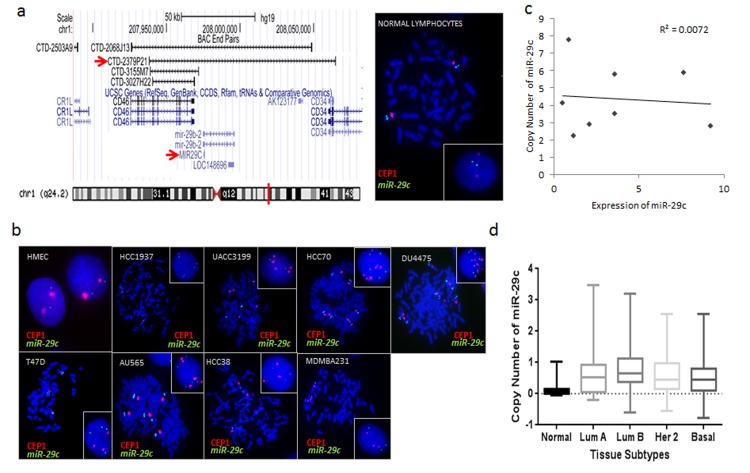
Fig 6. Fluorescent In Situ Hybridization (FISH) of miR-29c in breast cancer cell lines.
(a) Genomic position of BAC CTD-2379P21 at chromosome 1q32. This clone was selected for a homebrewed miR-29c FISH probe. Right image shows photomicrograph of miR-29c:CEP 1 FISH probes in control normal lymphocytes in metaphase and interphase (insert). (b) Photomicrographs of miR-29c:CEP1 in breast cancer cell lines are presented and show marked karyotype abnormalities. Cells were counterstained with DAPI (blue), while miR-29c is localized by green fluorescent signal, and CEP1 is localized by a red flourescent signal. Metaphase and interphase (insert) cells are shown. These Results are summarized in Table 1 and described further in S1 Text. (c) Scatter plot showing no correlation between copy number of miR-29c (from FISH) and expression of miR-29c in the breast cancer cell lines. (d) TCGA copy number data analysis confirms no significant difference in copy number of miR-29c between the different breast cancer subtypes in human tumors.

