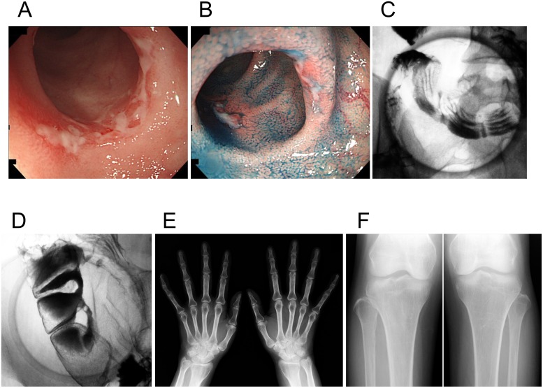
Fig 2. Clinical images of an individual with chronic nonspecific multiple ulcers of the small intestine (patient A-V–2).
(A, B) Retrograde ileoscopy shows active circular and oblique multiple ulcers with mucous exudates in the ileum. (C, D) A barium follow-through examination with compression shows multiple circular barium flecks (C), eccentric deformities, and strictures (D) in the ileum. (E, F) Radiographs of the hands and tibiofibulae show no obvious abnormalities such as cortical thickening of the metacarpals and periosteal hyperostosis.

