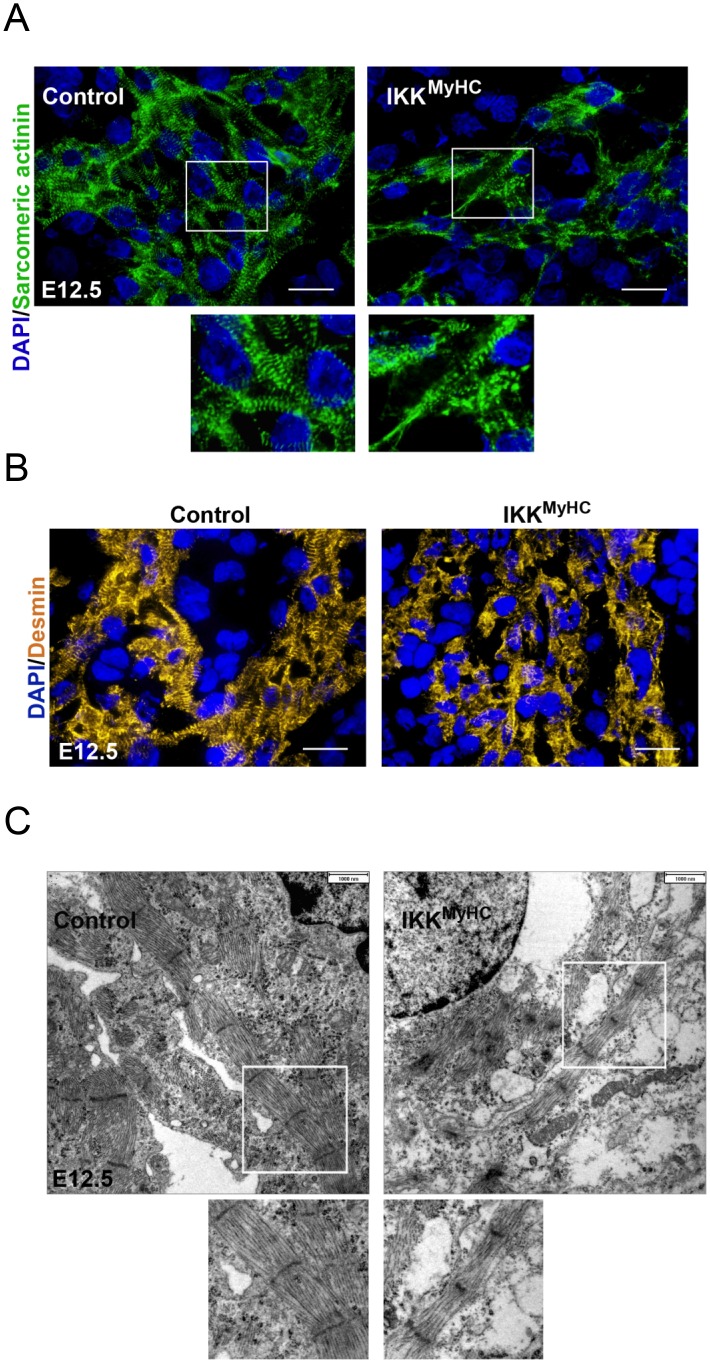
Fig 4. Cardiomyocyte-specific IKK/NF-κB activation induces myofiber degeneration and loss of typical cardiomyocyte proteins.
(A) Control and IKKMyHC sections were stained for sarcomeric α-actinin (green) and DAPI (blue). Scale bar: 100 μm. (B) Control and IKKMyHC sections were stained for desmin (yellow) and DAPI (blue). Scale bar: 100 μm. (C) Electron microscopic analyses of control and IKKMyHC heart sections. Cardiomyocytes of control animals show normally developed myofibers with typical sarcomeres (left). IKKMyHC cardiomyocytes show general disorganization and a reduced width of myofibers (right). Scale bar: 1 μm.

