
Fig 4. The lack of Tm7sf2 gene protects from LPS-induced acute kidney injury.
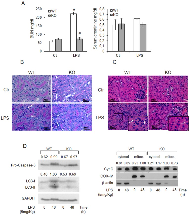
(A) WT and KO mice were i.p. injected with 5mg/Kg LPS and blood urea nitrogen (BUN) and creatinine were assessed after 24h. Results are given as mean ±s.d., (n = 5). *p<0.05 vs. control WT, # p<0.05 vs. the respective WT. Kidney sections of 72h LPS-treated WT and KO mice were stained with (B) periodic-reactive schiff (PAS) or with (C) hematoxylin/eosin staining. Arrows indicate interstitial oedema and vacuoles. Images magnification, x40.(D) Caspase 3, Cytocrome C, and LC3B in kidney after 48h of LPS-exposure. β-actin and GAPDH antibodies were used as loading control for total and cytosolic extracts. COX-IV was used as loading control for mitochondrial preparation.