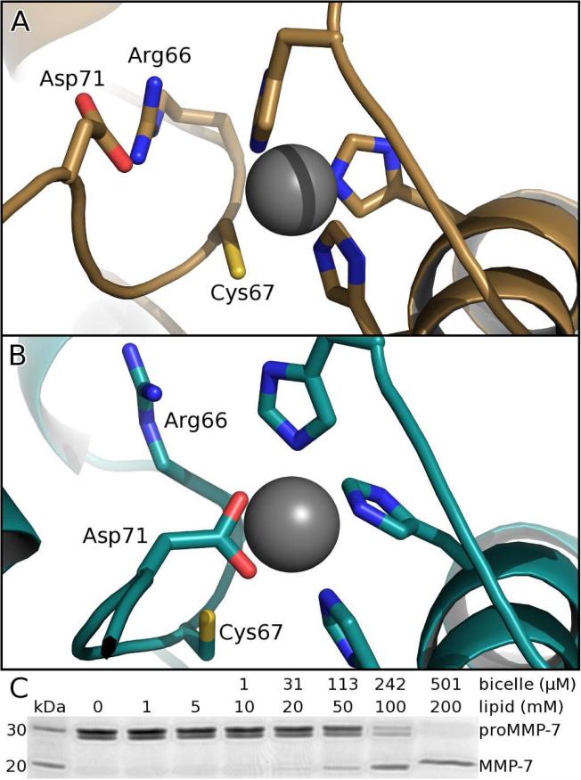
Figure 6. Bicelles alter auto-inhibitory conformation and stimulate activation.
(A) Free proMMP-7 features normal proximity of the catalytic zinc to the conserved cysteine of the auto-inhibitory segment of the pro-domain.
(B) When bound to DMPC/DH6PC bicelles, the conserved auto-inhibitory peptide switches to place the conserved aspartate at the zinc, according to the altered NOE patterns highlighted in Figure S6.
(C) DMPC/DH6PC bicelles (q=0.5) of sufficient concentration accelerate activation (2 h at 37°C) of proMMP-7 (7 μM) to mature MMP-7 (migrating faster by SDS-PAGE).

