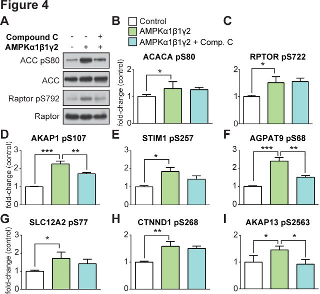
Fig. 4. AMPK substrate prediction using data-independent MS analysis of a global AMPK in vitro kinase assay.
(A) Immunoblot analysis of known AMPK substrates (ACC and Raptor) in HEK293 lysates subjected to a global AMPK (α1β1γ2) in vitro kinase assay ± active AMPK and the AMPK inhibitor Compound C. (B) – (I) Targeted quantification (mean ± standard deviation, one-way ANOVA corrected for multiple testing, *P < 0.05, **P < 0.01, ***P < 0.005, n=3) of phosphopeptides from HEK293 lysates subjected to a global AMPK (α1β1γ2) in vitro kinase assay using nanoUHPLC-MS/MS on a Q-Exactive MS operated in DIA.

