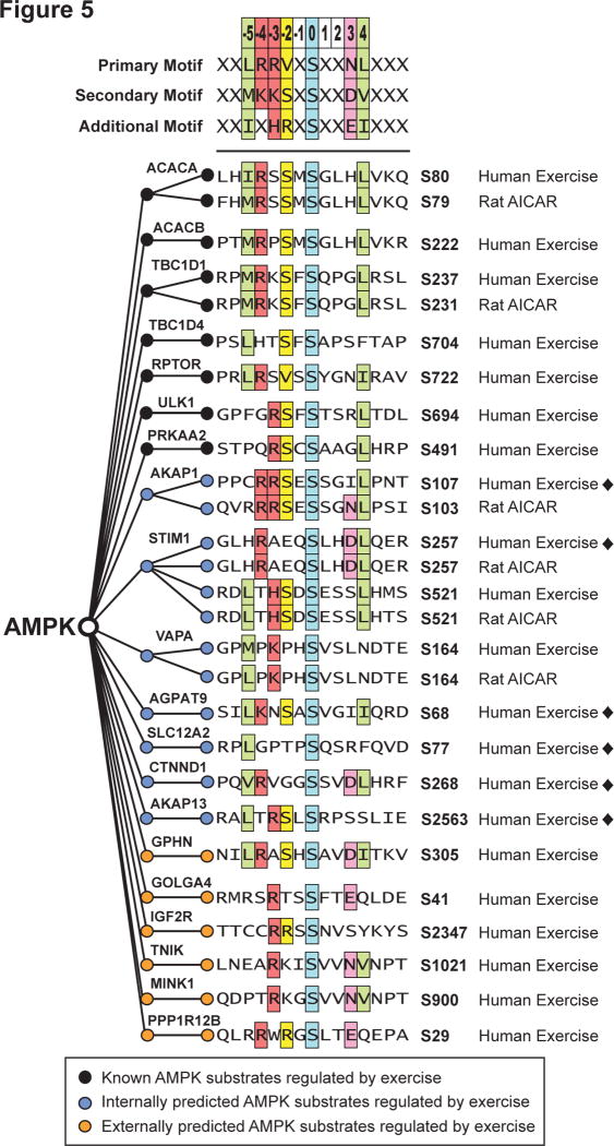
Fig. 5. AMPK consensus motif and substrate sequence alignment.
AMPK consensus motif derived from (Gwinn et al., 2008) with multiple sequence alignment of exercise-regulated phosphosites in human muscle (Human Exercise) is displayed. The alignment shows: known AMPK substrates (black circles); internally predicted AMPK substrates with validation (blue circles) in either AICAR stimulated Rat L6 myotubes (Rat AICAR) or global AMPK in vitro kinase analysis in HEK lysates (black diamonds ◆); and externally predicted AMPK substrates from (Banko et al., 2011) (orange circles).

