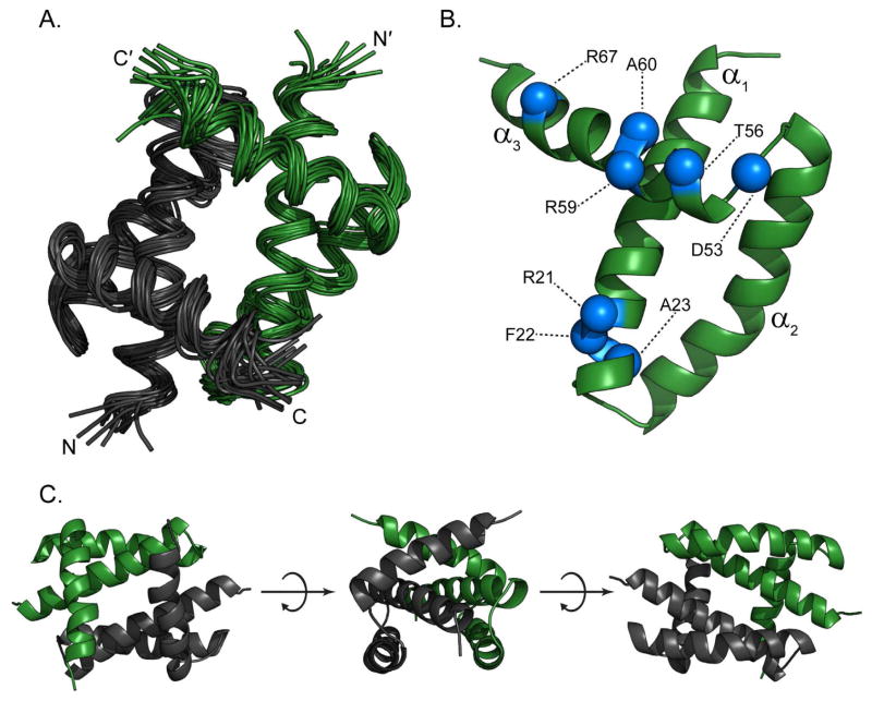
Figure 5. Solution NMR structural studies of 1918H1N1 NS1RBD.
(A) Ribbon diagram of the 16 energy-minimized conformers that represent the NMR structure of the 1918H1N1 NS1RBD (1–73) homodimer. The two monomers are shown in black and green respectively with the N- and C-termini labeled for each. (B) Monomeric structure with residues that are mutated when comparing the 1918H1N1 NS1RBD to the Udorn NS1RBD indicated in blue. (C) Multiple views of the 1918 H1N1 NS1RBD indicating that it retains the canonical six-helical fold demonstrated by previously solved solution structure of influenza Udorn NS1RBD.

