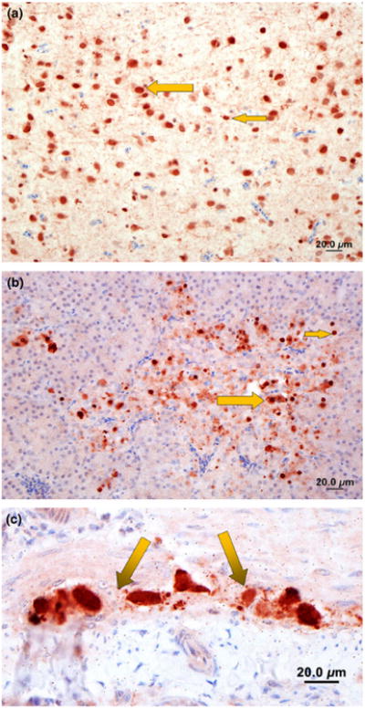
Fig. 5.

Immunohistochemistry staining for viral nucleoprotein (brown) of brain, pancreas and intestine of ducks infected with avian influenza A (H5N1) virus in Netrokona, Bangladesh, 2011. (a) Brain: moderate amount of nucleoprotein of avian influenza (AI) in the nucleus and cytoplasm of neurons (large arrow) and glial cells (small arrow). (b) Pancreas: large amount of AI antigen in the nucleus and cytoplasm of acinar cells (small arrow) and a few mononuclear inflammatory cells (large arrow) (c) Intestine: large amount of nucleoprotein of AI antigen in the nucleus and cytoplasm of subserosal and submucosal ganglia (arrows, myenteric plexus).
