Figure 2.

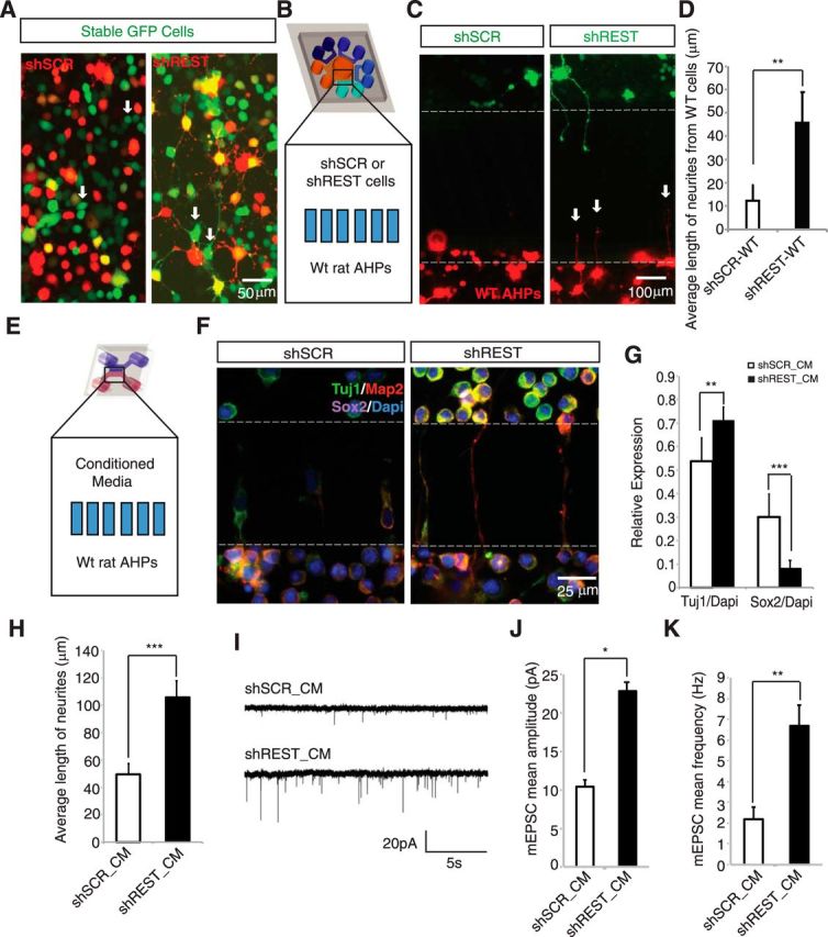
Conditioned medium from REST knockdown cells promote differentiation and maturation of AHPs non–cell-autonomously. A, A non–cell-autonomous effect was observed for REST knockdown cells. AHPs stably expressing GFP were transfected with shREST mCherry or shSCR mCherry plasmid. Electroporation efficiency was not 100%; thus, cells that did not take up the shRNA plasmids are green, whereas transfected cells are yellow. Green cells with neurites are seen in shREST but not in shSCR cultures (indicated by white arrows). B, Microfluidic compartmentalized coculture of either shSCR- or shREST-GFP transfected cells with wild-type (WT) cells-mCherry. Multicompartment design was introduced to see the non–cell-autonomous effect from shSCR and shREST toward the same population of WT cells simultaneously. C, Treatment chamber was plated with either shSCR- or shREST-GFP-transfected AHPs, and the response of WT AHPs (red cells) was monitored. WT AHPs showed neuronal morphology when the treatment chamber contained shREST-transfected cells (white arrows). D, Average length of processes from WT AHPs upon coculture with either shSCR or shREST cells (n = 12). **p < 0.005. E, Scheme showing two-chambered microfluidic device. In the top chamber (treatment chamber), we added conditioned medium from culture of shSCR- or shREST-transfected cells; WT AHPs were plated in the bottom chamber (cell chamber). F, shREST conditioned medium promotes differentiation of WT AHPs. Conditioned media were collected after 4 d of culture of shSCR- and shREST-transfected cells and then added to the treatment chamber for culturing WT AHPs (cell chamber). Map2-positive neurites (red) can be seen in the microgrooves in the shREST but not in the shSCR cultures. Green represents TUJ1. Magenta represents Sox2. Cells in the upper chamber (treatment chamber) result from migration through the microgrooves in both shSCR and shREST conditioned media. G, shREST conditioned medium (shREST_CM) caused an increase in TUJ1 expression (**p < 0.005, n = 24), whereas a reverse effect was observed for Sox2 expression (***p < 0.0001, n = 24). H, Average neurite length of AHPs cultured in conditioned medium taken from either shSCR- or shREST-transfected cells. A significant increase (***p < 0.0001, n = 50 for shREST_CM and n = 72 for shSCR_CM) in the average neurite length was observed in the treatment with shREST_CM in Map2(+) cells. I, Example of traces of mEPSCs recorded from primary neurons treated with conditioned medium from shSCR- or shREST-transfected cells. J, Treatment with shREST_CM led to a significant increase in mEPSC amplitude (mean 22.8 ± 4.3 pA, n = 10) compared with cells treated with shSCR_CM (mean 10.4 ± 0.8 pA, n = 8). *p < 0.05 (Student's t test). K, Cells treated with shREST_CM also exhibited an increase in mEPSC frequency (mean 6.7 ± 1.0 Hz) compared with shSCR_CM-treated cells (mean 2.2 ± 0.6 Hz). **p < 0.005 (Student's t test).
