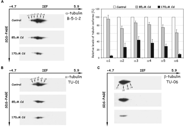
FIGURE 5.
Representative immunoblots probed with different antibodies against α-tubulin (A,B) and β-tubulin isoforms (C). Individual tubulin isoforms are denoted by arrows marked α1–α6 for α-tubulin and β1–β4 for β-tubulin. The quantitative results for α-tubulin (antibody B-5-1-2) were calculated as a ratio of pixel intensity values to area of spots and data were presented considering the control as a reference point (100%). The values represent the average of three independent measurements with a standard deviation.

