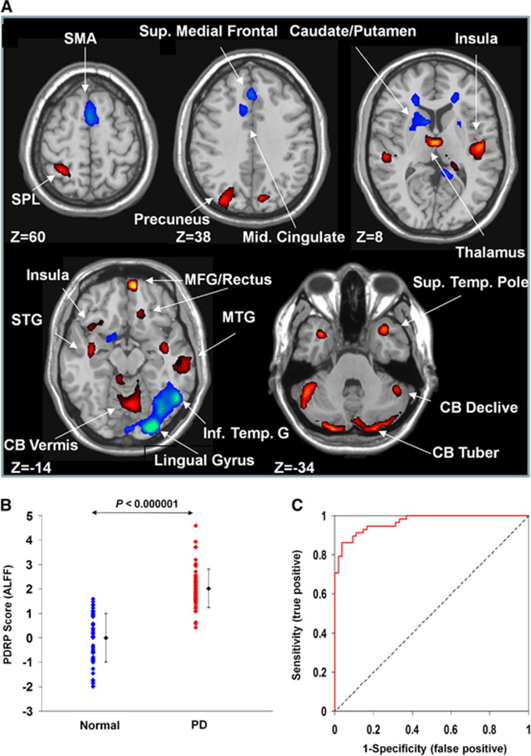
Figure 1.
Parkinson's disease–related spatial covariance pattern–amplitude of low-frequency fluctuation (PDRP–ALFF) identified with resting-state functional magnetic resonance imaging (fMRI). (A) The pattern was defined using ALFF images from 58 patients with PD and 54 normal controls in cohort A. This network topography was reliable at P<0.025 (see Table 1 for more information). (B) Group discrimination by PDRP–ALFF expression in patients with PD and normal controls. The expression of PDRP–ALFF significantly discriminated the PD patients from the normal controls (P<0.000001). (C) Receiver operating characteristic curve for discriminating the PD patients from the normal controls. The discriminatory power in cohort A was greater than that in cohort B or cohort C (see Figure 2D). The covariance pattern is superimposed on a standard T1-weighted MRI brain template and displayed with a height threshold of ±0.5 and an extent threshold of 30 voxels (240 mm3). Voxels with positive region weights (increased activity) are color-coded red and those with negative region weights (decreased activity) are color-coded blue. Error bars represent the s.d. plotted around the mean of each subject group. CB, cerebellum; MFG, medial frontal gyrus; MTG, middle temporal gyrus; SMA, supplementary motor area; SPL, superior parietal lobule; STG, superior temporal gyrus.

