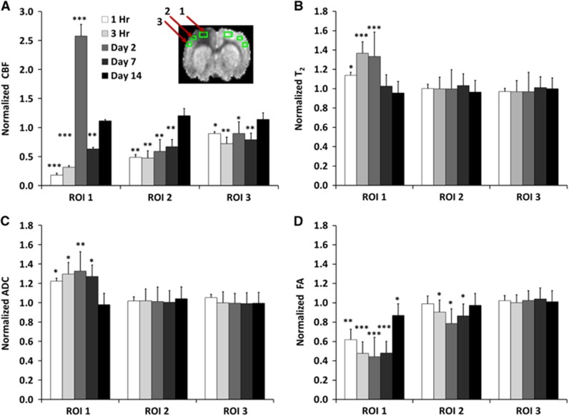
Figure 2.
Normalized (A) cerebral blood flow, (B) T2, (C) apparent diffusion coefficient, and (D) fractional anisotropy from the ipsilesional cortex at different time points after traumatic brain injury. Values were normalized to homologous region in the contralesional cortex (mean±s.e.m., n=8, * P<0.05, ** P<0.01, *** P<0.001 between ipsilesional and contralesional sides). Cerebral blood flow, T2, apparent diffusion coefficient, and fractional anisotropy of the homologous region in the contralesional cortex did not change with time and were not statistically different from sham-operated animals.

