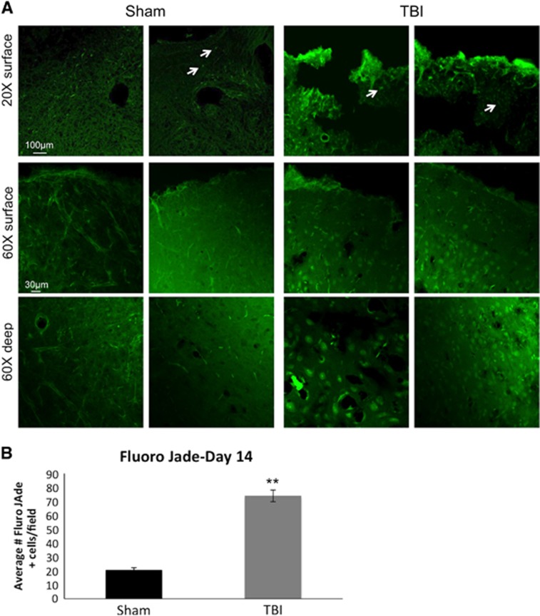
Figure 6.
(A) Representative images for Fluro Jade staining (green) are shown at × 20 and × 60 from the cortex of sham and traumatic brain injury animals. Positive Fluro Jade cells are indicated by white arrows. (B) Histogram demonstrating the average number of Fluro Jade positive cells within the cortex in sham and traumatic brain injury animals on day 14 post traumatic brain injury.

