Figure 4.
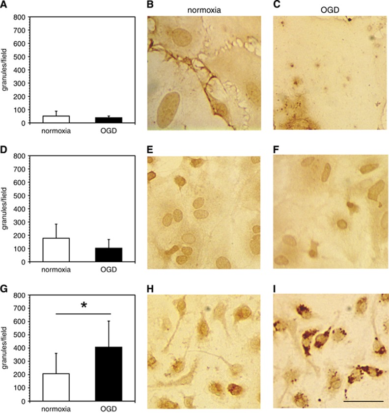
Association of immunoreactive cathepsin L with murine primary endothelial cells, astrocytes, and microglial cells. Immunoreactive cathepsin L is significantly associated with microglia under oxygen-glucose deprivation (OGD). Murine primary endothelial cells (A–C), astrocytes (D–F), and microglia (G–I) were cultured on 12-mm glass cover slips coated with poly-D-lysine (PDL) or collagen IV and subjected to normoxia or OGD. After fixation, the slides were immunostained with a rat anti-cathepsin L monoclonal antibody (MoAb) (see Supplementary Table 1). Photomicrographs are representative fields displaying the absence or presence of stained granules of cathepsin L associated with each cell type. In panels A, D, and G, data are shown as the number of granules/particles per microscopic field (× 40) (mean±s.d. from 10 fields each). There were no statistically significant differences in numbers of granules/particles per microscopic field under normoxia vs. OGD in endothelial cells (t18=1.01, P=0.33), or in astrocytes (t18=1.89, P=0.075). In comparison, numbers of granules/particles per microscopic field under normoxia were significantly reduced vs. OGD in microglia (t18=−2.55, P=0.02). Magnification bar=20 μm.

