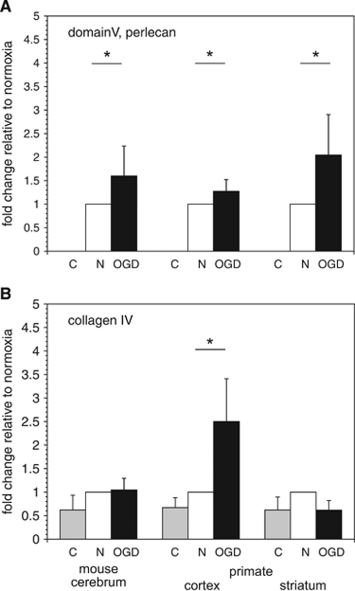
Figure 7.
Degradation of vascular extracellular matrix (ECM) by microglial secretion products. Degradation of perlecan domain V (26 kDa) (A) and the collagen IVα2 chain (167 kDa) (B) from murine cerebral cortex, and non-human primate cortex and striatum as shown by Western immunoblot. Tissue samples were incubated with microglia-conditioned medium ex vivo at 37°C for 2 hours and then processed for immunoblots. (A) Microglia subject to OGD produced a significant increase in perlecan degradation (increase in domain V release) from murine cerebral cortex (*, P=0.02; n=4), and from primate cortex (*, P=0.049; n=6) and striatum (*, P=0.003; n=6). (B) In contrast, microglia subject to OGD produced a significant increase only in collagen IV degradation from primate cortex only (*, P=0.003; n=6). Data shown as fold changes of tissue release in control buffer (C, shaded bars) and secretion products of microglia subject to OGD (filled bars) relative to those subject to normoxia (open bars). Data are from four samples in murine cortex, and six replicates of primate cortex and striatum. Each bar represents a separate experimental condition as displayed by the legend below the abscissa: C=vehicle, n=normoxia, and OGD=experimental ischemia in vitro.

