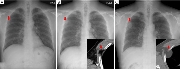
Figure 1.
(A) In the initial chest radiograph, a mass was detected in the right upper lung field (arrow); (B) 2 years after A, chest radiographs and CT scans showed a deep-seated intramuscular lipoma originating from the anterior serratus muscle (arrows); (C) 4 years after B, chest wall lipoma penetrated the third intercostal muscle and compressed the lung (arrows).

