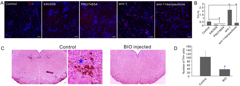
Figure 6. The effects of Wnt and GSK-3 inhibitor on mDA neuron differentiation.
(A) The effects of wnt signaling on the dopaminergic neuronal differentiation of ReNCell VM cells. Cells were treated with 1 μM XAV939, 2 μM PNU74654, 20 ng/ml human recombinant Wnt-1 protein, 3 μm kenpaullone or the indicated combination. Cells were stained with TH antibody. (B) Quantification of the percentage of TH+ cells after the treatments in A. In all the experiment control received equal volumes of DMSO treatment. (C) Injection of cell permeable GSK-3 inhibitor BIO significantly inhibited the development of TH+ dopamine neuron in the developing mouse midbrain. Left, an example slice of the control animals; middle, high resolution image of the *region in left image to confirm the TH+ neuron morphology; right, an example slice of the BIO injected animals. (D) Quantification of the number of TH+ cells in the midbrain slices. Numbers shown are the averages of 9 slices from 3 different animals. Scale bars, 50 μm. *P < 0.05 (red, significantly increased; blue, significantly decreased).

